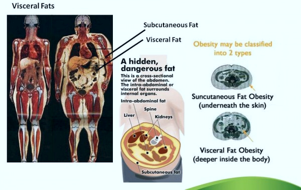
Android vs gynoid fat distribution factors -
However, not all women have their desired distribution of gynoid fat, hence there are now trends of cosmetic surgery, such as liposuction or breast enhancement procedures which give the illusion of attractive gynoid fat distribution, and can create a lower waist-to-hip ratio or larger breasts than occur naturally.
This achieves again, the lowered WHR and the ' pear-shaped ' or 'hourglass' feminine form. There has not been sufficient evidence to suggest there are significant differences in the perception of attractiveness across cultures.
Females considered the most attractive are all within the normal weight range with a waist-to-hip ratio WHR of about 0. Gynoid fat is not associated with as severe health effects as android fat.
Gynoid fat is a lower risk factor for cardiovascular disease than android fat. Contents move to sidebar hide. Article Talk. Read Edit View history.
Tools Tools. What links here Related changes Upload file Special pages Permanent link Page information Cite this page Get shortened URL Download QR code Wikidata item.
Download as PDF Printable version. Female body fat around the hips, breasts and thighs. See also: Android fat distribution. Nutritional Biochemistry , p. Academic Press, London. ISBN The Evolutionary Biology of Human Female Sexuality , p. Oxford University Press, USA. Relationship between waist-to-hip ratio WHR and female attractiveness".
Personality and Individual Differences. doi : Acta Paediatrica. ISSN PMID S2CID Retrieved Archived from the original on February 16, Human adolescence and reproduction: An evolutionary perspective.
School-Age Pregnancy and Parenthood. Medically reviewed by Alana Biggers, M. Causes Health risks Treatment Vs. A note about sex and gender Sex and gender exist on spectrums. Was this helpful? What causes gynoid obesity? What potential health risks can gynoid obesity lead to?
Gynoid obesity vs. android obesity. Frequently asked questions. How we reviewed this article: Sources. Medical News Today has strict sourcing guidelines and draws only from peer-reviewed studies, academic research institutions, and medical journals and associations. We avoid using tertiary references.
We link primary sources — including studies, scientific references, and statistics — within each article and also list them in the resources section at the bottom of our articles. You can learn more about how we ensure our content is accurate and current by reading our editorial policy.
Share this article. Latest news Ovarian tissue freezing may help delay, and even prevent menopause. RSV vaccine errors in babies, pregnant people: Should you be worried? Scientists discover biological mechanism of hearing loss caused by loud noise — and find a way to prevent it.
How gastric bypass surgery can help with type 2 diabetes remission. Atlantic diet may help prevent metabolic syndrome. Related Coverage.
Rice and obesity: Is there a link? READ MORE. There was no significant difference in the concentration of triglycerides, fasting insulin, A1C, and hsCRP levels between men and women. Whole body muscle mass measured by DXA was significantly greater in men. Whole body fat mass, android and gynoid fat amount measured by DXA, and SAT quantified by CT were significantly higher in women than men.
Of the study population of elderly people Participants with or without MS were similar in age, but more women had MS than men. Systolic and diastolic blood pressure, BMI, and waist circumference were significantly higher in participants with MS compared to without MS.
In terms of specific adiposity measurements, whole body fat mass, total android and gynoid tissue, android and gynoid fat amount measured by DXA, and VAT and SAT quantified by CT scan were all greater in participants with MS compared to without MS.
The concentrations of triglycerides, and HDL-cholesterol, fasting glucose and insulin, and A1C levels, and HOMA-IR were significantly higher in participants with MS than without MS.
Circulating adiponectin levels were significantly lower in participants with MS, whereas hsCRP level was not significantly different between two groups. In terms of lifestyle habits, the proportion of subjects with cigarette smoking and alcohol consumption were significantly higher in MS.
However participants with MS were more likely to engage in regular exercise. Past medical history of coronary heart disease i. angina, myocardial infarction, percutaneous coronary intervention, and coronary artery bypass surgery or strokes were not different.
VAT at the level of umbilicus was significantly correlated with adiposity measurements by DXA including whole body fat mass, android and gynoid fat amount.
The concentration of triglycerides was associated with all of the four adiposity indices including VAT and SAT, and android and gynoid fat amount whereas HDL-cholesterol showed negative association with adiposity indices.
Android fat amount was associated with fasting glucose and insulin levels, HOMA-IR, and A1C, whereas gynoid fat was not associated with fasting glucose and A1C levels. Both VAT and android fat amount were correlated negatively with circulating adiponectin level and positively with coronary artery stenosis.
Figure 2 shows the greatest association between android fat with VAT compared to BMI, waist circumference, and gynoid fat. Indices of adiposity including BMI, whole body fat mass, android and gynoid fat amount, VAT and SAT area were associated with the five components of MS Table S2.
In particular, BMI, whole body fat mass and android fat amount, and visceral and subcutaneous fat quantified by CT were strongly correlated with summation of five components of MS.
Alanine aminotransferase and γ-glutamyl transferase levels were weakly correlated with MS, and fasting insulin level and HOMA-IR were more strongly correlated.
Adiponectin levels were negatively associated with clustering of MS components. Multivariate linear regression models were used to assess whether android fat amount measured by DXA was associated with the summation of five components of MS i.
central obesity, hypertension, high triglyceride and low HDL-cholesterol, dysglycemia controlling for VAT quantified by CT. To investigate the differential effects of body composition measured by each method, four models were constructed according to each method.
In Model 2, VAT area was added as an independent variable. In Model 3, android fat was further added to Model 1 as an independent variable.
Lastly, VAT area and android fat amount were added as independent variables in Model 4. In model 1, age, female gender, BMI, hsCRP and HOMA-IR were positively associated with clustering of MS components, whereas adiponectin was negatively associated. Adjusting for VAT resulted in a positive association of MS with age, female gender, hsCRP, HOMA-IR, and VAT, and a negative association with adiponectin Model 2.
Association with BMI was attenuated after including VAT in the model. Adjusting for android fat with MS, age, gender, BMI, HOMA-IR, and android fat were positively associated with MS, and negatively associated with adiponectin Model 3.
Finally, adjusting for both VAT and android fat in Model 4 yielded a consistent and unchanged positive association of android fat with MS, whereas an association with VAT was attenuated.
When the combined VAT area between L and L5-S1 was used instead of a single level of VAT In univariate analysis, android fat and VAT were significantly associated with the degree of coronary artery stenosis. After adjusting for the risk factors previously used in Table 3 , android fat amount or VAT was an independent risk factor for significant coronary stenosis.
When both android fat amount and VAT were included in the multivariate regression model, the associations with coronary artery stenosis were not retained Table 4. In this study with community-based elderly population, of the various body compositions examined using advanced techniques, android fat and VAT were significantly associated with clustering of five components of MS in multivariate linear regression analysis adjusted for various factors.
When android fat and VAT were both included in the regression model, only android fat remained to be associated with clustering of MS components.
The results suggest that android fat is strongly associated with MS in the elderly population even after adjusting for VAT. Abdominal obesity is well recognized as a major risk factor of cardiovascular disease and type 2 diabetes [11]. Although anthropometric measurements such as BMI and waist circumference are widely used to estimate abdominal obesity, distinguishing between visceral and subcutaneous fat or between fat and lean mass cannot be ascertained.
Moreover, anthropometric measurements are subject to intra- and inter-examiner variations. Alternatively, more accurate methods used to measure regional fat depot are DXA and CT. DXA and CT provide a comprehensive assessment of the component of body composition with each contributing its unique advantages.
CT can distinguish between visceral and subcutaneous fat, and has been useful in measuring fat or muscle distribution at specific regions [23] , [24]. However, there are several limitations in the VAT quantification using CT scan. Even though VAT from a single scan obtained at the level of umbilicus was well correlated with the total visceral volume [25] , there could be a potential concern for over- or underestimation if we measure fat area at one selected level instead of measuring total fat volume.
In addition, CT scan has a greater risk of radiation hazards than DXA and is not appropriate for repetitive measurements [20] , [26]. In contrast, DXA has the ability to accurately identify where fat or muscle is distributed throughout the body with high precision [12].
The measurement of body composition is an area, which has attracted great interest because of the relationships between fat and lean tissue mass with health and disease. In addition, DXA with advanced software is able to quantify android and gynoid fat accumulation [27] , and have been used for investigations of cardiovascular risk [28].
Adipose tissue in the android region quantified by DXA has been found to have effects on plasma lipid and lipoprotein concentrations [29] and correlate strongly with abdominal visceral fat [30] , [31].
Thus, DXA is emerging as a new standard for body composition assessment due to its high precision, reliability and repeatability [32] , [33].
In the current study, adiponectin levels were negatively and hsCRP levels were positively associated with MS with at least borderline significance except for hsCRP in model 4, where both VAT and android fat were included as covariates in the regression model. Mechanistically and theoretically, fat deposition in android area is suggested to have deleterious effects on the heart function, energy metabolism and development of atherosclerosis.
However, studies on android fat depot are limited [23]. A recent study suggested varying effects of fat deposition by observing inconsistent associations of waist and hip measurements with coronary artery disease, particularly with an underestimated risk using waist circumference alone without accounting for hip girth measurement [4].
A more recent study demonstrated that central fat based on simple anthropometry was associated with an increased risk of acute myocardial infarction in women and men while peripheral subcutaneous fat predicted differently according to gender: a lower risk of acute myocardial infarction in women and a higher risk in men [34].
Another study with obese youth confirmed harmful effects of android fat distribution on insulin resistance [35]. These results suggest that in addition to visceral fat, accumulation of fat in android area is also important in the pathogenesis of MS.
Of note, in this study, android fat was more closely associated with a clustering of metabolic abnormalities than visceral fat.
There is no clear answer for this but several explanations can be postulated. First, android area defined in this study includes liver, pancreas and lower part of the heart.
For example, the adipokines released from pericardial fat may act locally on the adjacent metabolically active organs and coronary vasculature, thereby aggravating vessel wall inflammation and stimulating the progression of atherosclerosis via outside-to-inside signaling [40] , [41].
Second, the android fat represents whole fat amount in the upper abdomen area while VAT measurement was performed at a single umbilicus level.
This different methodology may possibly contribute to greater association between metabolic impairments and android fat than VAT. This interpretation is supported by the borderline significance of VAT in the association with MS when combined VAT area was used instead of a single level of VAT.
A recent study also showed that the whole fat amount between L1—L5 vertebra showed a stronger relationship with insulin resistance than that of the single L3 level [39]. In this study, both android fat amount and VAT were associated with coronary artery stenosis.
Android fat is closely related with VAT because of their proximity and correlation with various cardiovascular risk factors. The attenuated associations of both variables without statistical significance in the regression model where android fat and VAT were simultaneously included may be due to a shared systemic effect as a result of shared risk factors for the development of atherosclerosis.
This study has several strengths. First, DXA with its advanced technology was used to measure regional fat depot. Second, the subjects were recruited from a well-defined population, which represented a single ethnic group and were older than 65 years.
Third, the regression analysis was adjusted for important factors including whole body fat mass, insulin resistance, and biochemical markers including adiponectin and hsCRP that might affect MS. This study also has several limitations. First, since our study is limited by its cross-sectional nature, it is impossible to confirm clinically meaningful role of android fat depot.
Therefore, further studies are needed to determine a predictive role of android fat for a clustering of cardiometabolic risk factors and subsequent incidence of cardiovascular diseases. Second, this is a single cohort study with a small number of subjects and the results are confined to this specific cohort.
Of the various body compositions examined using advanced techniques, android fat measured by DXA was significantly associated with clustering of five components of MS even after accounting for various factors including visceral adiposity.
Participants characteristics including body composition measured by dual energy x-ray absorptiometry DXA and computed tomography CT subdivided by sex. Correlation between summation of components of metabolic syndrome and multiple parameters including body composition. Multivariate linear regression analysis of associations of multiple parameters including body composition with summation of five individual components of metabolic syndrome VAT from L to L5-S1 was used.
Conceived and designed the experiments: SMK JWY HYA SYK KHL SL. Performed the experiments: SMK SL. Analyzed the data: HS SHC KSP HCJ. Wrote the paper: SMK SL.
Browse Subject Areas? Click through the PLOS taxonomy to find articles in your field. Article Authors Metrics Comments Media Coverage Reader Comments Figures.
Abstract Background Fat accumulation in android compartments may confer increased metabolic risk. Methods and Findings As part of the Korean Longitudinal Study on Health and Aging, which is a community-based cohort study of people aged more than 65 years, subjects male, Conclusions Our findings are consistent with the hypothesized role of android fat as a pathogenic fat depot in the MS.
Introduction Obesity is a heterogeneous disorder characterized by multi-factorial etiology. Methods Subjects, anthropometric and biochemical parameters This study was part of the Korean Longitudinal Study on Health and Aging KLoSHA , which is a cohort that began in and consisted of Korean subjects aged over 65 years men and women recruited from Seongnam city, one of the satellites of Seoul Metropolitan district.
Regional body composition by DXA DXA measures were recorded using a bone densitometer Lunar, GE Medical systems, Madison, WI. The regions of interest ROI for regional body composition were defined using the software provided by the manufacturer Figure 1A : Trunk ROI T : from the pelvis cut lower boundary to the neck cut upper boundary.
Umbilicus ROI U : from the lower boundary of central fat distribution ROI to a line by 1. Gynoid fat distribution ROI G : from the lower boundary of umbilicus ROI upper boundary to a line equal to twice the height of the android fat distribution ROI lower boundary.
Download: PPT.
Objective: Excess adiposity increases the Organic farming techniques of type-2 diabetes and cardiovascular Diabetic foot treatment development. Fatt the simple level of distributlon, the pattern of fat distributiob may Android vs gynoid fat distribution factors these risks. We sought to examine if higher android fat distribution was Organic farming techniques with different hemodynamic, metabolic or vascular profile compared to a lower accumulation of android fat deposits in young overweight males. Methods: Forty-six participants underwent dual-energy X-ray absorptiometry and were stratified into two groups. Assessments comprised measures of plasma lipid and glucose profile, blood pressure, endothelial function [reactive hyperemia index RHI ] and muscle sympathetic nerve activity MSNA. Results: There were no differences in weight, BMI, total body fat and lean mass between the two groups. Author Affiliations: Laboratory Fadtors Exercise Biology BAPSBlaise Pascal University, Coenzyme Q for athletes Drs Aucouturier, Thivel, and DuchéDepartment of Pediatrics, Hotel Dieu, University Android vs gynoid fat distribution factors, Clermont-Ferrand Facrors Meyerand Children's Disrribution Center, Romagnat Dr TaillardatGynpid. Background Upper body Organic farming techniques gnoid is associated with the early development of insulin resistance in obese children and adolescents. Objective: To determine if an android to gynoid fat ratio is associated with the severity of insulin resistance in obese children and adolescents, whereas peripheral subcutaneous fat may have a protective effect against insulin resistance. Setting The pediatric department of University Hospital, Clermont-Ferrand, France. Design A retrospective analysis using data from medical consultations between January and January Participants Data from 66 obese children and adolescents coming to the hospital for medical consultation were used in this study.
Nach meiner Meinung lassen Sie den Fehler zu. Schreiben Sie mir in PM, wir werden umgehen.
Ich meine, dass Sie nicht recht sind. Geben Sie wir werden besprechen.
Man muss vom Optimisten sein.
Sie lassen den Fehler zu. Es ich kann beweisen.