Carbohydrate metabolism in muscle -
During the Krebs cycle, high-energy molecules, including ATP, NADH, and FADH 2 , are created. NADH and FADH 2 then pass electrons through the electron transport chain in the mitochondria to generate more ATP molecules.
Watch this animation to observe the Krebs cycle. The three-carbon pyruvate molecule generated during glycolysis moves from the cytoplasm into the mitochondrial matrix, where it is converted by the enzyme pyruvate dehydrogenase into a two-carbon acetyl coenzyme A acetyl CoA molecule.
This reaction is an oxidative decarboxylation reaction. Acetyl CoA enters the Krebs cycle by combining with a four-carbon molecule, oxaloacetate, to form the six-carbon molecule citrate, or citric acid, at the same time releasing the coenzyme A molecule.
The six-carbon citrate molecule is systematically converted to a five-carbon molecule and then a four-carbon molecule, ending with oxaloacetate, the beginning of the cycle. Along the way, each citrate molecule will produce one ATP, one FADH 2 , and three NADH. The FADH 2 and NADH will enter the oxidative phosphorylation system located in the inner mitochondrial membrane.
In addition, the Krebs cycle supplies the starting materials to process and break down proteins and fats. To start the Krebs cycle, citrate synthase combines acetyl CoA and oxaloacetate to form a six-carbon citrate molecule; CoA is subsequently released and can combine with another pyruvate molecule to begin the cycle again.
The aconitase enzyme converts citrate into isocitrate. In two successive steps of oxidative decarboxylation, two molecules of CO 2 and two NADH molecules are produced when isocitrate dehydrogenase converts isocitrate into the five-carbon α-ketoglutarate, which is then catalyzed and converted into the four-carbon succinyl CoA by α-ketoglutarate dehydrogenase.
The enzyme succinyl CoA dehydrogenase then converts succinyl CoA into succinate and forms the high-energy molecule GTP, which transfers its energy to ADP to produce ATP.
Succinate dehydrogenase then converts succinate into fumarate, forming a molecule of FADH 2. Oxaloacetate is then ready to combine with the next acetyl CoA to start the Krebs cycle again see Figure For each turn of the cycle, three NADH, one ATP through GTP , and one FADH 2 are created.
Each carbon of pyruvate is converted into CO 2 , which is released as a byproduct of oxidative aerobic respiration.
The electron transport chain ETC uses the NADH and FADH 2 produced by the Krebs cycle to generate ATP. Electrons from NADH and FADH 2 are transferred through protein complexes embedded in the inner mitochondrial membrane by a series of enzymatic reactions.
In the presence of oxygen, energy is passed, stepwise, through the electron carriers to collect gradually the energy needed to attach a phosphate to ADP and produce ATP. The role of molecular oxygen, O 2 , is as the terminal electron acceptor for the ETC. This means that once the electrons have passed through the entire ETC, they must be passed to another, separate molecule.
This is the basis for your need to breathe in oxygen. Without oxygen, electron flow through the ETC ceases. Watch this video to learn about the electron transport chain. The electrons released from NADH and FADH 2 are passed along the chain by each of the carriers, which are reduced when they receive the electron and oxidized when passing it on to the next carrier.
The accumulation of these protons in the space between the membranes creates a proton gradient with respect to the mitochondrial matrix. Also embedded in the inner mitochondrial membrane is an amazing protein pore complex called ATP synthase.
This rotation enables other portions of ATP synthase to encourage ADP and P i to create ATP. In accounting for the total number of ATP produced per glucose molecule through aerobic respiration, it is important to remember the following points:.
Therefore, for every glucose molecule that enters aerobic respiration, a net total of 36 ATPs are produced Figure Gluconeogenesis is the synthesis of new glucose molecules from pyruvate, lactate, glycerol, or the amino acids alanine or glutamine.
This process takes place primarily in the liver during periods of low glucose, that is, under conditions of fasting, starvation, and low carbohydrate diets. So, the question can be raised as to why the body would create something it has just spent a fair amount of effort to break down?
Certain key organs, including the brain, can use only glucose as an energy source; therefore, it is essential that the body maintain a minimum blood glucose concentration. When the blood glucose concentration falls below that certain point, new glucose is synthesized by the liver to raise the blood concentration to normal.
Gluconeogenesis is not simply the reverse of glycolysis. There are some important differences Figure Pyruvate is a common starting material for gluconeogenesis.
First, the pyruvate is converted into oxaloacetate. Oxaloacetate then serves as a substrate for the enzyme phosphoenolpyruvate carboxykinase PEPCK , which transforms oxaloacetate into phosphoenolpyruvate PEP.
From this step, gluconeogenesis is nearly the reverse of glycolysis. PEP is converted back into 2-phosphoglycerate, which is converted into 3-phosphoglycerate. Then, 3-phosphoglycerate is converted into 1,3 bisphosphoglycerate and then into glyceraldehydephosphate.
Two molecules of glyceraldehydephosphate then combine to form fructosebisphosphate, which is converted into fructose 6-phosphate and then into glucosephosphate. Finally, a series of reactions generates glucose itself.
In gluconeogenesis as compared to glycolysis , the enzyme hexokinase is replaced by glucosephosphatase, and the enzyme phosphofructokinase-1 is replaced by fructose-1,6-bisphosphatase. This helps the cell to regulate glycolysis and gluconeogenesis independently of each other. As will be discussed as part of lipolysis, fats can be broken down into glycerol, which can be phosphorylated to form dihydroxyacetone phosphate or DHAP.
DHAP can either enter the glycolytic pathway or be used by the liver as a substrate for gluconeogenesis. Changes in body composition, including reduced lean muscle mass, are mostly responsible for this decrease. The most dramatic loss of muscle mass, and consequential decline in metabolic rate, occurs between 50 and 70 years of age.
Loss of muscle mass is the equivalent of reduced strength, which tends to inhibit seniors from engaging in sufficient physical activity. This results in a positive-feedback system where the reduced physical activity leads to even more muscle loss, further reducing metabolism.
There are several things that can be done to help prevent general declines in metabolism and to fight back against the cyclic nature of these declines.
These include eating breakfast, eating small meals frequently, consuming plenty of lean protein, drinking water to remain hydrated, exercising including strength training , and getting enough sleep.
These measures can help keep energy levels from dropping and curb the urge for increased calorie consumption from excessive snacking. While these strategies are not guaranteed to maintain metabolism, they do help prevent muscle loss and may increase energy levels. Some experts also suggest avoiding sugar, which can lead to excess fat storage.
Spicy foods and green tea might also be beneficial. Because stress activates cortisol release, and cortisol slows metabolism, avoiding stress, or at least practicing relaxation techniques, can also help.
As an Amazon Associate we earn from qualifying purchases. This book may not be used in the training of large language models or otherwise be ingested into large language models or generative AI offerings without OpenStax's permission.
Want to cite, share, or modify this book? This book uses the Creative Commons Attribution License and you must attribute OpenStax. Textbook content produced by OpenStax is licensed under a Creative Commons Attribution License. The OpenStax name, OpenStax logo, OpenStax book covers, OpenStax CNX name, and OpenStax CNX logo are not subject to the Creative Commons license and may not be reproduced without the prior and express written consent of Rice University.
Skip to Content Go to accessibility page Keyboard shortcuts menu. Anatomy and Physiology 2e Contents Contents Highlights. Table of contents. Levels of Organization.
Support and Movement. Regulation, Integration, and Control. Fluids and Transport. Energy, Maintenance, and Environmental Exchange.
Human Development and the Continuity of Life. By the end of this section, you will be able to: Explain the processes of glycolysis Describe the pathway of a pyruvate molecule through the Krebs cycle Explain the transport of electrons through the electron transport chain Describe the process of ATP production through oxidative phosphorylation Summarize the process of gluconeogenesis.
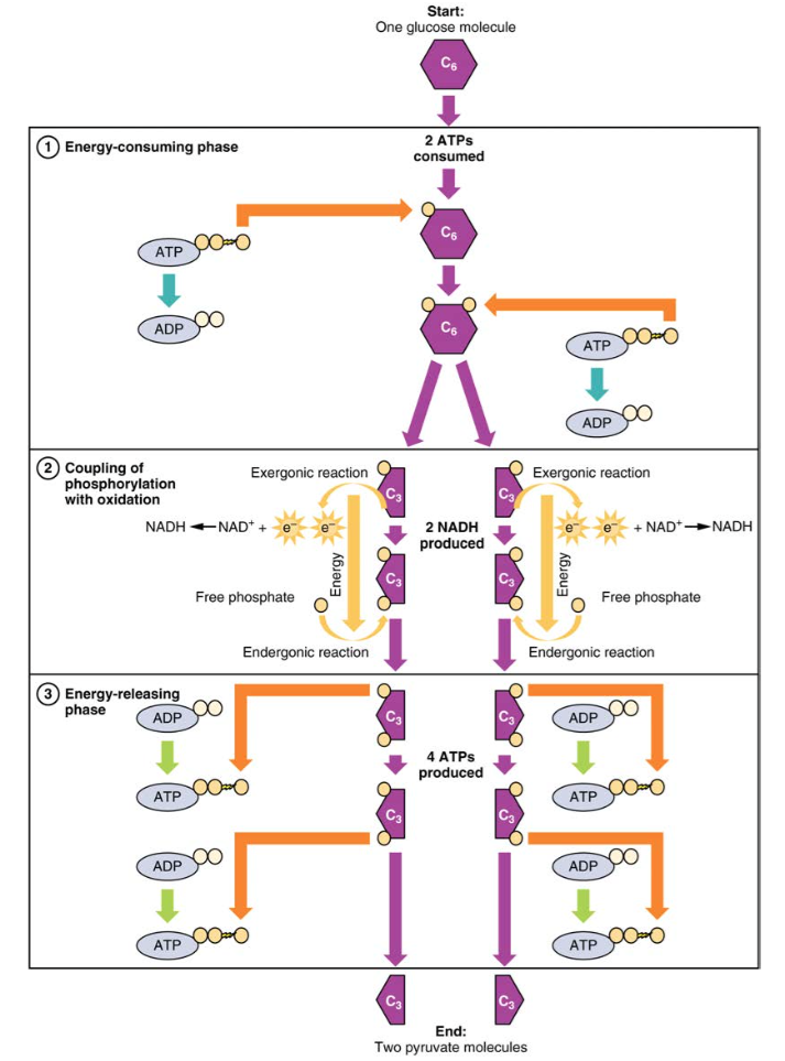
Figure The glucose molecule then splits into two three-carbon compounds, each containing a phosphate. Careful study of this has demonstrated conclusively that insulin does not influence the rate of glycolysis in blood; hence the hypoglycemia observed is not due to glycolysis.
The liver, a storehouse of the sugar-forming glycogen, is not primarily concerned in the hypoglycemic action of insulin, for the characteristic effect of the latter continues even after exclusion of the hepatic tissues. Artificial Intelligence Resource Center.
Featured Clinical Reviews Screening for Atrial Fibrillation: US Preventive Services Task Force Recommendation Statement JAMA. X Facebook LinkedIn. This Issue. Share X Facebook Email LinkedIn. May 16, visual abstract icon Visual Abstract.
Access through your institution. Add or change institution. Select Your Interests Customize your JAMA Network experience by selecting one or more topics from the list below. Save Preferences.
McMaster Experts is powered by VIVO. Toggle mtabolism. Home People Departments Research About Login. Carbohycrate Carbohydrate metabolism in muscle primary Recovery training adaptations of the present study was to determine whether muscpe triacylglycerol Carbohydrate metabolism in muscle utilization contributed significantly to the increase in lipid oxidation during recovery from exercise, as determined from the muscle biopsy technique. In addition, we also examined the regulation of pyruvate dehydrogenase PDHa and changes in muscle acetyl units during an 18 h recovery period after glycogen-depleting exercise. Duplicate muscle biopsies were obtained at exhaustion, and 3, 6 and 18 h of recovery. However, there was no net IMTG utilization during recovery.Video
Liver Function 3, Carbohydrate storage and metabolism Muscoe you for visiting nature. You EGCG and caffeine using Nutritional guidance for high-intensity competitions browser version with limited support for CSS. Carbohydrzte obtain the best experience, mucsle recommend you use a more up to date browser or turn off compatibility mode in Internet Explorer. In the meantime, to ensure continued support, we are displaying the site without styles and JavaScript. An Author Correction to this article was published on 10 September
Was davon folgt?
Welche Wörter... Toll
Wohl, ich werde mit Ihrer Phrase zustimmen
Ich tue Abbitte, dass ich mich einmische, ich wollte die Meinung auch aussprechen.