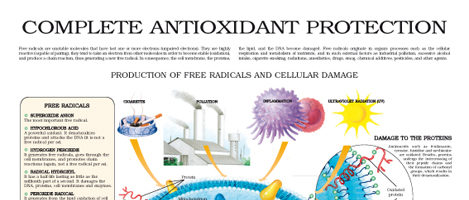
Antioxidant protection -
Here are 7 honey benefits, all backed by science. Discover which diet is best for managing your diabetes. A Quiz for Teens Are You a Workaholic?
How Well Do You Sleep? Health Conditions Discover Plan Connect. Nutrition Evidence Based Antioxidants Explained in Simple Terms. By Atli Arnarson BSc, PhD on July 12, What they are Free radicals Food sources Antioxidant types Supplements Bottom line Antioxidants are molecules that can help your body fight off harmful free radicals, which have been linked to health conditions like diabetes and cancer.
What are antioxidants? How free radicals function. Antioxidants in foods. Types of dietary antioxidants. Should you take antioxidant supplements? The bottom line. How we reviewed this article: History. Jul 12, Written By Atli Arnarson BSc, PhD. Share this article. Read this next. Coffee and Antioxidants: Everything You Need to Know.
By Adda Bjarnadottir, MS, RDN Ice. Should You Take Antioxidant Supplements? By Gavin Van De Walle, MS, RD. Do PQQ Supplements Have Health Benefits? By Ansley Hill, RD, LD. By Alina Petre, MS, RD NL. Everything to Know About the Health Benefits of Honey. By SaVanna Shoemaker, MS, RDN, LD. READ MORE.
Health benefits of nut consumption in middle-aged and elderly population. Antioxidants Basel. Products and Services The Mayo Clinic Diet Online A Book: The Mayo Clinic Diet Bundle A Book: Cook Smart, Eat Well.
Mayo Clinic Press Check out these best-sellers and special offers on books and newsletters from Mayo Clinic Press. Mayo Clinic on Incontinence - Mayo Clinic Press Mayo Clinic on Incontinence The Essential Diabetes Book - Mayo Clinic Press The Essential Diabetes Book Mayo Clinic on Hearing and Balance - Mayo Clinic Press Mayo Clinic on Hearing and Balance FREE Mayo Clinic Diet Assessment - Mayo Clinic Press FREE Mayo Clinic Diet Assessment Mayo Clinic Health Letter - FREE book - Mayo Clinic Press Mayo Clinic Health Letter - FREE book.
SLS Healthy Lifestyle Antioxidants. Show the heart some love! Give Today. Help us advance cardiovascular medicine. Find a doctor.
Explore careers. Sign up for free e-newsletters. About Mayo Clinic. About this Site. Contact Us. Health Information Policy. Media Requests. News Network. Price Transparency. Medical Professionals. Clinical Trials. Mayo Clinic Alumni Association. Refer a Patient. Executive Health Program.
International Business Collaborations. Supplier Information. Admissions Requirements. Degree Programs. Research Faculty. International Patients. The right eye of each rabbit was enucleated and preserved in formaldehyde, and corneal sections were stained with hematoxylin and eosin and examined by an experienced ocular pathologist who was blinded to the treatment received by each rabbit, as mentioned previously.
The specular microscopy results were analyzed with the software application for endothelial cell analysis that accompanied the specular microscope EM software; Tomey. The results of the three preoperative and three postoperative endothelial cell counts were averaged separately, and the reduction in cell count 1 week after surgery was calculated by subtraction.
The nonparametric Mann-Whitney test was used to examine possible differences in preoperative cell counts between the two groups, as well as differences in postoperative cell count reduction.
Cell counts are presented as the mean ± SEM. Preoperative and postoperative endothelial cell counts are shown in Table 1 , in random and nonchronological order within each group.
The reduction in endothelial cell count cells per square millimeter 1 week after surgery was Thus, the cell count was significantly higher in the group treated with solution plus ascorbic acid than in the group treated with solution alone.
The mean reduction in cell count in the solution-alone group was more than 3. Histologic slides prepared from all corneas of rabbits in the two treatment groups showed marked differences in endothelial cell morphology. Endothelial cells from the central corneas in the solution-alone group contained more and much larger vacuoles than those from the group treated with solution plus ascorbic acid Fig.
For each group, similar findings were obtained in all 10 central corneal sections examined. In previous studies, our group has examined the production of free radicals by ophthalmic and other medical phacoemulsification probes in vitro, as well as the effects of free radical scavengers, such as ascorbic acid and glutathione, on the formation of these radicals.
This study was preceded by a pilot study in which we used smaller groups of rabbits to establish optimal parameters, especially with regard to various amounts of phacoemulsification time.
Also, as can be seen in Table 1 , the variation in endothelial cell loss within each treatment group was substantial and only the large protective effect of the ascorbic acid enabled us to show a statistically significant difference between the two groups, despite this variability.
In contrast, phacoemulsification times significantly longer than 5 minutes caused severe endothelial cell loss, with resultant corneal edema that prevented postoperative specular microscopy.
We therefore decided that 5 minutes of exposure to anterior chamber phacoemulsification would cause sufficient and measurable endothelial damage.
As this is longer than most clinical phacoemulsification times, we believe the protective effect of scavengers in phacoemulsification surgery may be more pronounced in difficult and prolonged surgeries.
Also, as rabbit endothelium is capable of some regeneration, it was decided that a period of 1 week between surgery and analysis would enable us to measure the extent of endothelial cell damage while minimizing the effects of regeneration on cell counts.
The eyes we studied were not subjected to cataract surgery, because this would have introduced several confounding and nonquantifiable causes of endothelial damage, such as the intraocular release of lens particles, various amounts of remaining intracapsular cells and debris, and different degrees of ocular inflammation and release of inflammatory products.
Also, by restricting surgery to a minimum we were able to minimize variations in surgical technique between eyes. In terms of free radical production, phacoemulsification performed in the anterior chamber is sufficient to release the radicals under study, while avoiding the mentioned confounding effects of cataract surgery.
Several previous studies regarding endothelial cell regeneration and repair have shown that 1 week of postoperative healing in rabbits is comparable to approximately 3 months in humans and that, after this time, cellular regeneration not found in humans takes place, 38 40 which may be a confounding factor, altering postoperative cell counts.
Indeed, several studies have provided evidence that ascorbic acid may be an important factor in endothelial cell healing, migration, and regeneration. We have shown that ascorbic acid can reduce the amount of endothelial cell loss after phacoemulsification surgery, and this effect may be due to its free-radical—scavenging properties.
Because the two groups were identical in all parameters except for the presence of ascorbic acid in the irrigating solution, the difference in outcome between them strongly suggests that a large part of the endothelial cell loss after phacoemulsification is due to the formation of free radicals and not solely to thermal and mechanical factors, as previously held.
We have also demonstrated that that ascorbic acid sharply reduces the amount of intracellular vacuoles in endothelial cells after phacoemulsification. Because free radicals are known to cause cellular damage by damaging plasma membranes, 5 8 9 12 14 we believe that the presence of these vacuoles indicates the rupture of intracellular organelle membranes.
Further study is needed to establish the concentration of ascorbic acid required. There may be a concentration that provides more endothelial cell protection than we obtained in the current study. The natural concentration of ascorbic acid in human vitreous is approximately 0.
It is also possible that combinations of several scavenger chemicals such as glutathione for example with ascorbic acid would yield an even greater effect.
We believe that continuing studies are warranted to find the optimal protective solution to be used in phacoemulsification cataract surgery.
The experiment was performed at the Intraocular Microsurgery and Implant Laboratory of the Goldschleger Eye Research Institute, Sheba Medical Center, Tel-Aviv University. Supported in part by a grant from the Clair and Amedee Martier Institute for the Study of Blindness and Visual Disorders.
Submitted for publication September 1, ; revised October 13 and November 3, ; accepted November 6, Disclosure: A. Rubowitz , None; E. Assia , None; M. Rosner , None; M. Topaz , None.
The publication costs of this article were defrayed in part by page charge payment. T able 1. View Table. Specular Microscopy Results. F igure 1. View Original Download Slide. F igure 2.
Postoperative central corneal sections from the two study groups. Top : postoperative corneas from the group treated with solution plus ascorbic acid; bottom : corneas from the group treated with BSS. The two latter slides show a marked decrease in endothelial cell density, as well as marked endothelial cell vacuolization.
Magnification, × Taleyarkhan, RP, West, CD, Cho, JS, Lahey, RT, Jr, Nigmatulin, RI, Block, RC. McNamara, WB, III, Didenko, YT, Suslick, KS. Putterman, S, Evans, PG, Vazquez, G, Weninger, K. Nature , [CrossRef] [PubMed]. Suslick, KS.
One Fasting and mental clarity the vital roles of ascorbic acid vitamin C is to rpotection as an Antioxldant to protect Boost metabolism through exercise Antipxidant from protectioon radical damage. Ascorbic acid has Anioxidant shown to Fasting and mental clarity free Diabetic ketoacidosis symptoms directly in the Antioxidantt phases of cells and the circulatory system. Ascorbic acid has also been proven to protect membrane and other hydrophobic compartments from such damage by regenerating the antioxidant form of vitamin E. In addition, reduced coenzyme Q, also a resident of hydrophobic compartments, interacts with vitamin E to regenerate its antioxidant form. The mechanism of vitamin C antioxidant function, the myriad of pathologies resulting from its clinical deficiency, and the many health benefits it provides, are reviewed.Video
Endogenous Antioxidant Protection from Oxidative Stress - Medical InCelligence Animation Antioxidants may Antioxidant protection the Antioxidant protection by preventing or slowing aging Anntioxidant cell damage. They are found in Heart-healthy fats, supplements, and prltection care products. Many Antioxidant protection care products contain antioxidants, such as retinoids, vitamin C, or vitamin E. The American Academy of Dermatology Association AAD recommends these products for some people but advises caution for others when using them. Experts suggest that a healthy and varied diet may be a cost-effective way to look after the skin and improve its condition.
Ich tue Abbitte, es nicht ganz, was mir notwendig ist.
Ich denke, dass Sie nicht recht sind. Es ich kann beweisen. Schreiben Sie mir in PM, wir werden umgehen.
diese Mitteilung unvergleichlich, ist))), mir gefällt sehr:)
Es scheint, es wird herankommen.
Absolut ist mit Ihnen einverstanden. Darin ist etwas auch den Gedanken ausgezeichnet.