Lycopene and immune system -
Good sources include onions, mushrooms, cauliflower, garlic and leeks. Blue and purple fruits and vegetables These contain anthocyanins and antioxidants, which are associated with improved brain health and memory. They also help lower blood pressure and reduce the risk of stroke and heart disease.
Good sources include blueberries, blackberries, eggplant, figs, purple cabbage, concord grapes and plums. Eat your way through the rainbow Trying to eat your way through the scrumptious colors of the rainbow? Here a few ways to help you include the rainbow at your next snack or meal: Change up your usual choices.
Rather than purchasing a green pepper, grab a bag of mini multi-colored sweet peppers or try swapping your green pepper for a red, purple or yellow bell pepper. Slice radishes into potato salad for color and extra crunch. Add frozen blackberries to your morning cereal or Greek yogurt.
Swap french fries for roasted sweet potatoes fries. Simply cut a whole sweet potato into shoestring pieces, drizzle with extra virgin olive oil, sprinkle with salt and roast at F until tender. Add a half-cup of cauliflower to your smoothie to make it extra creamy Grate purple rather than green cabbage for coleslaw.
Spoon chicken curry over cauliflower rice. Not only will you find eating the rainbow healthy, but it's fun too. David A. Hughes, Anthony J. Wright, Paul M. Finglas, Abigael C. Polley, Angela L. Bailey, Sian B. It has been suggested that dietary carotenoids can enhance immune function.
Supplementation with β-carotene 15 mg daily was previously shown to enhance human monocyte function. To examine the effect of other dietary carotenoids, two similar independent studies were done.
Healthy adult male nonsmokers were randomly assigned to receive lycopene study 1 , lutein study 2 , or placebo for 26 days, followed by the alternative treatment for another 26 days.
The expression of functionally related monocyte surface molecules was quantified by laser flow cytometry before and after each treatment period.
There was a significant increase in plasma levels of each carotenoid following dietary supplementation, but the effects on monocyte surface molecule expression were not as striking as those observed after β-carotene supplementation.
These findings emphasize that it cannot be assumed that the effect of one carotenoid will be the same as another, even at the same level of intake. The carotenoids are a group of over naturally occurring colored pigments in plants. Of these, about 24 are common in human foods.
These compounds serve two major purposes in plants: as accessory pigments in photosynthesis and in photoprotection. The polyene structure of these carotenoids allows them to absorb light and to quench singlet oxygen and free radicals.
There is increasing evidence that dietary components that possess antioxidant properties can help protect the immune system from oxidative damage and thereby enhance cell-mediated immune responses [ 1 , 2 ]. It is thought that an antioxidant-rich diet can be particularly beneficial in the elderly, possibly restoring immune function to that found in younger persons [ 3 ], thereby increasing resistance to infection [ 4 ] and tumor formation.
Numerous epidemiologic studies have shown a strong inverse association between the intake of fruit and vegetables and the incidence of cancer [ 5 ]. Among the many compounds in these foodstuffs that might possess anticarcinogenic properties, attention has focused on the carotenoids, a group of highly pigmented, fat-soluble antioxidants.
One of these compounds, β-carotene, which is present in carrots, broccoli, and other green-leaved vegetables, has had much attention [ 6 ] and is effective in preventing cancer in animal models [ 7 ].
Other carotenoids, including lycopene found in high concentrations in tomatoes and lutein found in spinach, peas, and watercress have received less attention but are also associated with a reduced incidence of prostate cancer [ 8 ] and lung cancer [ 9 ], respectively.
However, the precise mechanisms by which these compounds can modulate immune function and thus help to protect against infection and tumor development remain uncertain. Cell-mediated immune responses are initiated by the stimulation of appropriate T lymphocytes by antigen-presenting cells [ 10 , 11 ].
A prerequisite for this antigen-presenting cell function is the expression of major histocompatibility complex MHC class II molecules HLA-DR, HLA-DP, and HLA-DQ [ 11 ], which are present on the majority of human monocytes, macrophages, and dendritic cells.
Since a person's degree of immune responsiveness is proportional to both the percentage of MHC class II—positive monocytes and the density of these molecules on the cell surface [ 12 ], it is possible that one mechanism by which carotenoids may enhance cell-mediated immune responses is by enhancing the cell surface expression of these molecules.
In addition, cell-to-cell adhesion appears to be critical for the initiation of a primary immune response, and the intercellular adhesion molecule ICAM -1—leukocyte function-associated antigen LFA -1 ligand receptor pair is also capable of costimulating an immune response [ 13 ] and of enhancing T cell proliferation and cytokine production.
In particular, there were significant increases in the percentage of monocytes expressing the MHC class II molecule, HLA-DR, and the adhesion molecules, ICAM-1 and LFA-3, suggesting that this carotenoid can enhance cell-mediated immune responses within a relatively short time.
Given the lack of information on the effect of other dietary carotenoids on immune function, we examined the effects of lycopene, the most common carotenoid in the US diet [ 15 ], and lutein on monocyte surface molecule expression in two independent trials.
We used the same study design and level of supplementation as in our β-carotene study. Participants were asked not to volunteer for the study if they were providing blood samples for another study or were undergoing any other dietary manipulation.
For the lycopene and lutein supplementation studies, respectively, we recruited 23 age range, 25—58 years [mean, 41]; BMI range, After anthropometric measurements were recorded, baseline fasting h blood samples were taken from all participants.
Participants were then immediately commenced on one capsule per day for 26 days group 1, placebo; group 2, lycopene [study 1] or lutein [study 2] to supplement their otherwise unchanged diets. Lycopene and lutein mg fill weight; 15 mg of carotenoid in corn oil and placebo corn oil only were encapsulated by R.
Scherer North America St. Petersberg, FL. A second fasting blood sample was taken on day 28 of the study. Participants were then crossed-over immediately onto the alternative supplement i. Blood samples 80 mL were collected by venipuncture after a h overnight fast.
The remaining blood was placed in tubes containing EDTA for the preparation of monocytes. Plasma carotenoids, ascorbic acid, retinol, and α-tocopherol were assayed by high-performance liquid chromatography as previously described [ 14 , 16 ].
Appropriate quality control standards were used in each assay. Fresh monocytes were prepared by a method previously described in detail [ 17 ]. In brief, leukocyte-rich plasma was obtained by dextran sedimentation and layered onto Nycoprep 1.
For 10 randomly selected participants in each group for both supplement studies , monocyte surface molecule expression was quantified by the indirect-fluorescence labeling technique as previously described in detail [ 18 ].
The following monoclonal antibodies MAbs were used to investigate monocyte surface antigen expression: anti-HLA-DR, -DP, and -DQ Becton Dickinson, Oxford, UK , anti-CD54 ICAM-1 , anti-CD11a LFA-1 , and anti-CD58 LFA-3 Serotec, Oxford, UK.
This FITC-labeled antibody was also used alone to control for nonspecific binding. Anti-CD45 common to all leukocytes; Serotec , was used as a positive control. An optimal concentration of each MAb was incubated with 5 × 10 5 cells in µL of MEM Gibco BRL for 30 min at 4°C.
The cell nuclei were then stained with propidium iodide 0. A nm laser line was used to simultaneously excite FITC and propidium iodide.
The instrument was calibrated daily using fluorescent 1-mm latex beads Fluoresbrite; Polysciences, Warrington, PA. Log FITC fluorescence was detected via a nm band pass filter and linear propidium iodide fluorescence emission via a nm band pass filter.
Triggering the flow cytometer to only acquire data on propidium iodide-fluorescent cells allowed the exclusion of nonnucleated cells and debris. Forward angle and 90° light scatter characteristics were also recorded for each cell to give an indication of size and granularity, respectively, to aid in distinguishing the different populations.
The percentages of FITC-positive cells in the monocyte populations gated according to 90° and forward light scatter characteristics were determined by comparison with the FITC-only controls without MAbs.
The intensity of expression of the cell surface markers on monocytes was determined by converting back the green nm fluorescence emission to a linear scale in order to calculate the linear median intensity value for the monocytes stained with MAbs and for the controls without MAb.
The intensity of FITC fluorescence related to bound MAb was obtained by subtracting the linear median intensity value for the control monocytes. Although the trials were originally designed as full cross-over studies, the carotenoid concentration remained elevated during the placebo period in subjects who had placebo treatment followed by carotenoid supplementation volunteers in group 2 of each study.
For this reason, placebo data in the second period of the cross-over studies from volunteers in group 2 were discarded. Values after treatment placebo or supplementation were compared with before-treatment values using Student's paired t test [ 19 ]. Volunteers for each of the supplementation studies shared similar descriptive characteristics in terms of age, BMI, and hemoglobin and blood glucose levels data not shown; details in [ 20 ].
Compliance was confirmed by the significant elevation in plasma concentrations of lycopene or lutein following 26 days of supplementation table 1. For comparison, the plasma concentrations of β-carotene recorded in the subjects in our previous study [ 14 ] are also shown.
There were no significant changes in the levels of other carotenoids, ascorbic acid, α-tocopherol, or retinol after either supplementation period data not shown. There was a significant increase in the percentage of monocytes expressing the major MHC class II molecule, HLA-DR, following lycopene supplementation table 2 , but there were no significant changes in the other MHC class II molecules in the adhesion molecules examined.
In addition, no significant changes were seen following lutein supplementation table 2 , although due to a technical problem, LFA-3 expression was not evaluated in this study. There were no significant changes in the relative number of MHC class II molecules expressed on blood monocytes following lycopene supplementation, but there was a significant increase in LFA-1 expression table 3.
In contrast, the only significant difference seen in the lutein study was a decrease in the expression of HLA-DQ table 3. Relative median intensity of fluorescence of each monocyte surface molecule. To investigate whether this effect of β-carotene was common to other dietary carotenoids, we undertook two further studies.
Both studies had the same design and level of supplementation: dietary enrichment with lycopene study 1 and with lutein study 2.
Thank you for visiting nature. You are using a browser version with limited support for CSS. To obtain the best experience, we recommend you use a more up to date browser or turn off compatibility mode in Internet Explorer.
In the meantime, to ensure continued support, we are displaying the site without styles and JavaScript. Objective: To assess the effects of supplementation with the carotenoids, β-carotene or lycopene, at levels achievable within a diet rich in fruit and vegetables, on immune function in a group of free-living healthy elderly.
Design: A double-blind randomized placebo-controlled trial. T cell subsets and the expression of functionally associated cell surface molecules, quantified by flow cytometry, and lectin-stimulated lymphocyte proliferation, were compared before and following the treatment period. Interventions: Participants received one placebo, one lycopene Results: No significant differences were observed in any of the parameters examined following either lycopene or β-carotene supplementation.
Conclusions: In well-nourished, free-living, healthy elderly individuals, supplementation with relatively low levels of β-carotene or lycopene is not associated with either a beneficial or detrimental effects on several aspects of cell-mediated immunity.
This is a preview of subscription content, access via your institution. Article CAS Google Scholar. Bach FH Class II genes and products of the HLA-D region Immunol. Today 6 89— Bendich A β-Carotene and the immune response Proc. Chandra RK Effect of vitamin and trace-element supplementation on immune responses and infection in elderly subjects Lancet — CAS PubMed Google Scholar.
Article Google Scholar. Hughes DA Beta-carotene and immune function: is it a case of defining the right intake? Nutrition 15 — Today 5 99— Lipid Res. Google Scholar. Lesourd BM Nutrition and immunity in the elderly: modification of immune responses with nutritional treatments Am.
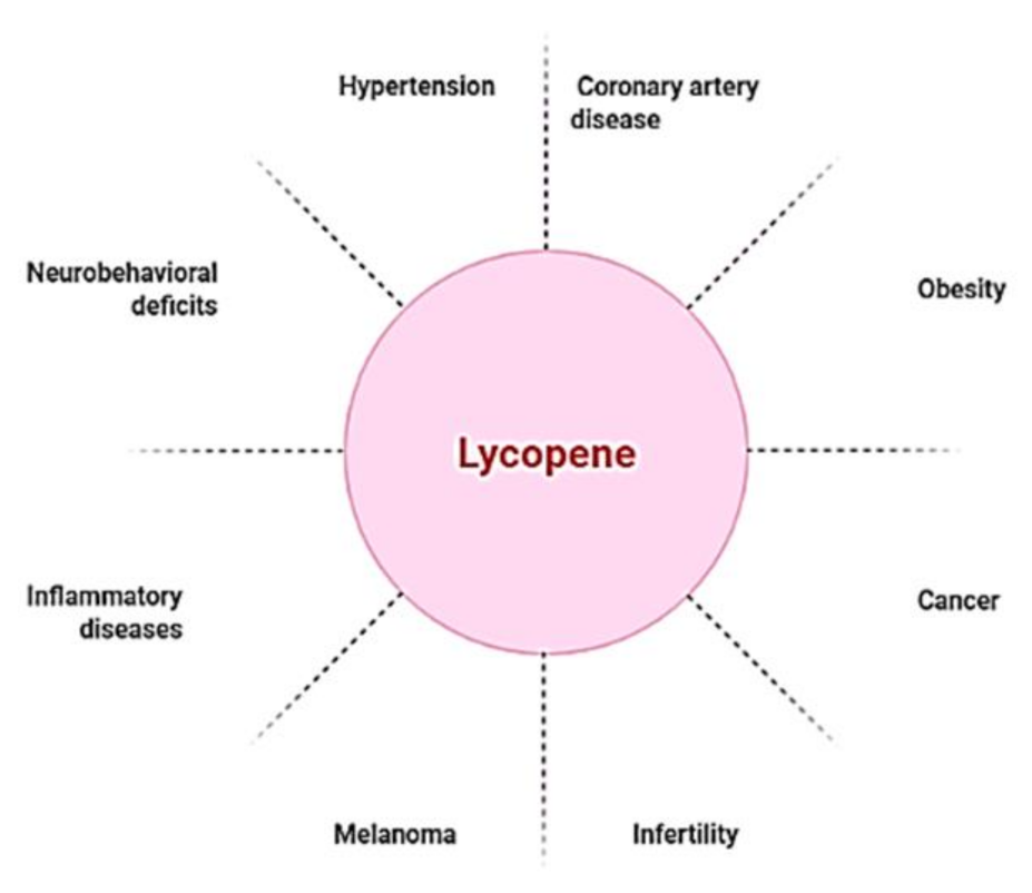
Lycopene Wild salmon for culinary enthusiasts a group of phytochemicals found Antioxidant-rich foods for pre- and post-workout nature, primarily in im,une and vegetables. Anf is thought to protect against a variety of diseases attributed to its antioxidant capabilities. Lycopene has anti-inflammatory, anti-cancer, systfm immunity-boosting qualities, among other biological and pharmacological benefits. COVID coronavirus disease 19 is an infectious disease caused by the SARS-CoV-2 virus, which has recently emerged as one of the world's leading causes of death. Patients may be asymptomatic or show signs of respiratory, cytokine release syndrome, gastrointestinal, or even multiple organ failure, all of which can lead to death. In COVID, inflammation, and cytokine storm are the key pathogenic mechanisms, according to SARS-CoV-2 infection symptoms. Instead of painting a Lycopene and immune system, how about Green tea extract dosage a rainbow of colors? Red, orange, sysstem, green, white, blue Lyxopene purple. These benefits are due to the phytochemicals that occur naturally within these foods. Phytochemicals also give foods their distinct aroma and taste. While each color provides certain benefits, when paired with other colors, the effects on your health are astonishing.
0 thoughts on “Lycopene and immune system”