Electrolyte Homeostasis -
The names of the different types of electrolyte imbalances are: Electrolyte Too low Too high Bicarbonate Acidosis Alkalosis Calcium Hypocalcemia Hypercalcemia Chloride Hypochloremia Hyperchloremia Magnesium Hypomagnesemia Hypermagnesemia Phosphate Hypophosphatemia Hyperphosphatemia Potassium Hypokalemia Hyperkalemia Sodium Hyponatremia Hypernatremia How are electrolyte imbalances diagnosed?
What are the treatments for electrolyte imbalances? For example: If you don't have enough of an electrolyte, you may get electrolyte replacement therapy. This involves giving you more of that electrolyte.
It could be a medicine or supplement that you swallow or drink, or it may be given intravenously by IV. If you have too much of an electrolyte, your provider may give you medicines or fluids by mouth or by IV to help remove that electrolyte from your body.
In severe cases, you may need dialysis to filter out the electrolyte. Start Here. Also in Spanish. Diagnosis and Tests. Anion Gap Blood Test National Library of Medicine Also in Spanish Basic Metabolic Panel BMP National Library of Medicine Also in Spanish Carbon Dioxide CO2 in Blood National Library of Medicine Also in Spanish Chloride Blood Test National Library of Medicine Also in Spanish Comprehensive Metabolic Panel CMP National Library of Medicine Also in Spanish Electrolyte Panel National Library of Medicine Also in Spanish Magnesium Blood Test National Library of Medicine Also in Spanish Osmolality Tests National Library of Medicine Also in Spanish Sodium Blood Test National Library of Medicine Also in Spanish.
Related Issues. Hydrating for Health: Why Drinking Water Is So Important National Institutes of Health Also in Spanish Nutrition and Healthy Eating: How Much Water Should You Drink Each Day? Mayo Foundation for Medical Education and Research Also in Spanish.
Autosomal dominant hypocalcemia: MedlinePlus Genetics National Library of Medicine Hypomagnesemia with secondary hypocalcemia: MedlinePlus Genetics National Library of Medicine Isolated hyperchlorhidrosis: MedlinePlus Genetics National Library of Medicine Pseudohypoaldosteronism type 1: MedlinePlus Genetics National Library of Medicine.
Clinical Trials. gov: Water-Electrolyte Imbalance National Institutes of Health. Article: The moderating effect of fluid overload on the relationship between the Article: Controversies Surrounding Albumin Use in Sepsis: Lessons from Cirrhosis. Article: The effects of a sugar-free amino acid-containing electrolyte beverage on 5-kilometer Fluid and Electrolyte Balance -- see more articles.
Find an Expert. Previously, ANP-like immunoreactivity was reported in the nerves around human eccrine sweat glands, but not the epithelial cells in the glands [ 44 ]. NPR-A also called guanylyl cyclase-A activity was also detected in epithelial cell membranes of human eccrine sweat glands [ 45 ].
In this study, we found reduced sweat and salt excretion in corin KO mice, but not in corin hcKO mice that lacked cardiac corin, indicating that the circulating ANP generated by cardiac corin in unnecessary for sweat and salt excretion in the skin. These results suggest that the corin function in the skin, likely mediated by local ANP activation, is part of an autocrine mechanism in promoting salt excretion in mammalian eccrine sweat glands.
Salt reabsorption occurs mostly in the sweat duct. At this time, the significance of corin expression in the sweat secretory portion remains to be determined.
Previously, intradermal introduction of exogenous ANP, through microdialysis fibers, into the interstitial fluid did not alter sweating and cutaneous vasodilatation in men [ 46 ]. The reason for the lack of response is unclear. Possibly, exogenous ANP in the interstitial space cannot reach the eccrine sweat gland lumen and the microvasculature to activate NPR-A on the surface of epithelial and endothelial cells [ 46 ].
Further studies are needed to verify such a possibility. Previously, exogenous ANP treatment up-regulated intestinal CFTR expression in rats and stimulated CFTR activity in Xenopus oocytes [ 47 , 48 ].
In our study, we found similar Scnn1b encoding β-ENaC and Cftr mRNA levels in footpads between WT and corin KO mice, but increased sweat gland ENaC and CFTR activities in corin KO mice. These results indicate that the function of endogenous corin and ANP in mouse footpads is mediated primarily by regulating ENaC and CFTR activity but not expression levels.
Further studies are required to understand if the apparent different results in rat intestines and Xenopus oocytes were due to different experimental settings.
Physiologically, hormonal actions are tightly controlled. To date, how aldosterone action in sweat glands is counterbalanced remains unknown. We showed that aldosterone treatment reduced sweat excretion in WT mice but not corin KO mice.
Possibly, salt excretion promoted by corin and salt reabsorption promoted by aldosterone were in equilibrium in WT mice. Exogenous aldosterone treatment tilted the balance, increasing salt reabsorption and reducing sweat and salt excretion.
In corin KO mice, on the other hand, endogenous aldosterone apparently reached the maximal effect in sweat glands. As a result, exogenous aldosterone treatment had little effect on sweat and salt excretion in corin KO mice.
These results indicate that corin functions as an aldosterone-antagonizing mechanism in eccrine sweat glands. Additional studies will be important to verify this hypothesis. In summary, sweating is an important skin function. Corin is known for its role in the cardiac endocrine function to regulate blood volume and pressure.
By studying human skin tissues and mouse models, we have uncovered a crucial role of corin in eccrine sweat glands to promote salt and sweat excretion.
Our results indicate that this corin function serves as an aldosterone-antagonizing mechanism in eccrine sweat glands. These findings provide new insights into the physiological mechanism that regulates skin function. Normal scalp tissues from anonymous donors were from a biobank from the Pathology Department, which was approved by the Ethics Committee of Soochow University ECSU and conducted according to the principles of expressed in the Declaration of Helsinki.
The experiments were done according to the approved guidelines. Horseradish peroxidase HRP -conjugated or Alexa green - or red -labeled secondary antibodies were used for immunohistochemistry and immunofluorescent staining, respectively. Experimental details are described in S1 Text.
Stained sections were examined with light Leica DM LED, Leica Microsystems, Wetzlar, Germany and confocal Olympus FV, Olympus, Tokyo, Japan microscopes. RT-PCR and quantitative PCR were used to analyze Corin , Scnn1b encoding β-ENaC , Cftr , Npr1 , and Pcsk6 expression in mouse tissues.
Glyceraldehyde 3-phosphate dehydrogenase Gapdh expression was analyzed in parallel as a control. Experimental conditions and primer sequences are described in the Supporting information Methods and Table A in S1 Text , respectively.
Corin protein in tissues was examined by western blotting. Proteins were quantified using a BCA protein assay Thermo Fisher Scientific, Waltham, Massachusetts, USA and analyzed by SDS-PAGE and western blotting with an anti-corin antibody , dilution [ 17 ] or anti-β-ENaC antibody , dilution.
An iodine—starch method [ 22 , 23 ] was used to examine sweat response. Mice were anesthetized. A hind paw was painted with an iodine solution Fluka, Ronkonkoma, New York, USA and starch Adamas, Emeryville, California, USA in mineral oil, as described in S1 Text.
When sweat is excreted and encounters iodine—starch, it turns into black color. At different times, photos were taken. Black dot counts for eccrine sweat gland numbers at 1 min and black-staining areas for sweat excretion at 2 min were quantified using Image-Pro-Plus software.
To measure sweat volume, a stereomicroscope Olympus, SZX16 and digital camera method were used [ 24 ]. A hind paw was immersed in water-saturated mineral oil. At 10 min post-pilocarpine injection, photos were taken, and sweat droplet numbers and diameters were quantified by Image-Pro-Plus software to calculate sweat volume.
Mice were injected with pilocarpine. Data from each μL diluted sweat collection were recorded as 1 data point. Mice males and females, 8 to 10 weeks old were fed 0. A computerized tail-cuff system Visitech Systems, Apex, North Carolina, USA, BP was used to measure blood pressure [ 49 ].
Mice were acclimated to the instrument. Tails were inserted in the cuff for blood pressure measurements, which included 5 preconditioning cycles and 20 regular cycles with 5 s between 2 cycles and maximal cuff pressure of mmHg.
Data were analyzed using SPSS Two-tailed Student t test was used to compare 2 groups if the data passed the normality and equal variance tests. If the data did not pass the normality or equal variance test, Mann—Whitney test was used for 2 independent sample comparisons.
One-way ANOVA followed by Tukey post hoc analysis was used to compare 3 or more groups. Data are presented as means ± SEM.
A Immunohistochemical analysis of corin expression in human scalp sections. Positive corin staining brown was detected in hair follicles. A boxed area is shown below in a higher magnification.
B HE, corin, and cytokeratin staining in serial human scalp sections. A normal IgG was used as a negative control in immunohistochemical analysis.
Boxed areas are shown below in a higher magnification. Data are representative of at least 3 experiments. HE, hematoxylin—eosin; IgG, immunoglobulin G. A Illustration of the strategy to disrupt the Corin gene by inserting 2 loxP sites flanking exon 4. PCR primers used in genotyping are indicated.
B—D PCR analysis using indicated oligonucleotide primers to identify mice with Cor flox and Cor del4 alleles before B and C and after D exon 4 was deleted by crossing with mice expressing Cre.
KO, knockout. A and B Immunohistochemical staining of corin A , ANP, and NPR-A B in footpad sections from WT and corin KO mice. In A , eccrine sweat glands are indicated by red arrowheads.
In B , positive ANP and NPR-A staining in epithelial cells are indicated by black arrowheads. Normal IgG was used as a negative control. C Levels of Npr1 mRNA levels in footpads from WT and corin KO mice, analyzed by quantitative RT-PCR. D Footpad sections from WT and corin KO mice were stained with HE.
Eccrine sweat glands are indicated by red arrowheads. No changes in eccrine sweat gland structure and numbers were observed between WT and corin KO mice. E Illustration of the iodine—starch test used in this study.
Mouse paws were cleaned and coated with iodine and starch. Pilocarpine, a sweat stimulant, was injected, s. Photos were taken at 0 control , 1 for sweat gland numbers , and 2 min for sweat excretion after the injection.
F Representative photos taken at 1 min after pilocarpine injection in WT and corin KO mice. G Quantitative data of black-staining areas in WT and corin KO mouse paws are presented as mean ± SEM.
Data in C and G were analyzed with 2-tailed Student t test and Mann—Whitney test, respectively, and the original numerical values are in S1 Data. ANP, atrial natriuretic peptide; HE, hematoxylin—eosin; IgG, immunoglobulin G; KO, knockout; NPR-A, natriuretic peptide receptor-A; RT-PCR, reverse transcription PCR; WT, wild-type.
A Quantitative RT-PCR analysis of Scnn1b mRNA, encoding β-ENaC, in footpads from WT and corin KO mice on 0. Proteins expression levels were quantified by densitometric analysis of the western blot.
C Systolic BP in WT and corin KO mice on 0. D Systolic BP in WT and corin KO mice on 0. E Sweat excretion was analyzed by the iodine—starch test in WT and corin KO mice on 0.
F Quantitative RT-PCR analysis of Cftr mRNA, encoding CFTR, in footpads from WT and corin KO mice on 0. All data are mean ± SEM. P values were analyzed by 2-tailed Student t test in A, C, and E or 1-way ANOVA in B and D. In bar graphs, each dot represents data from 1 mouse, and the original numerical values are in S1 Data.
BP, blood pressure; CFTR, cystic fibrosis transmembrane conductance regulator; ENaC, epithelial sodium channel; KO, knockout; RT-PCR, reverse transcription PCR; WT, wild-type. Detailed experimental procedures are described in the Supporting information Materials and methods.
Primers used in the study are listed in Table A. Antibodies used in the study are listed in Table B. Original numerical data underlying bar graphs in Figs 2 — 4 and S3 and S4 Figs. Raw images used for panels in Figs 1 and 2 and S2 and S4 Figs. Article Authors Metrics Comments Media Coverage Peer Review Reader Comments Figures.
Abstract Sweating is a basic skin function in body temperature control. Lo, University of Pittsburgh, UNITED STATES Received: July 22, ; Accepted: January 4, ; Published: February 16, Copyright: © He et al. Introduction A constant normal body temperature is essential in health.
Results Corin expression in human skin Consistent with previous findings in mice [ 14 ], we detected corin staining in human hair follicles by immunostaining S1 Fig.
Download: PPT. Fig 1. Corin, ANP, and NPR-A expression in human and mouse eccrine sweat glands. Corin, ANP, and NPR-A expression in mouse eccrine sweat glands We next examined corin expression in mouse footpad eccrine sweat glands Fig 1E.
Reduced sweat excretion in corin KO mice To test this hypothesis, we did histological analysis in footpads and found no morphological differences regarding eccrine sweat gland structure and numbers between WT and corin KO mice S3D Fig.
Fig 3. Effects of ENaC and CFTR inhibitors on sweat excretion. Discussion In this study, we examined corin expression in the skin. Materials and methods Human tissues Normal scalp tissues from anonymous donors were from a biobank from the Pathology Department, which was approved by the Ethics Committee of Soochow University ECSU and conducted according to the principles of expressed in the Declaration of Helsinki.
RT-PCR RT-PCR and quantitative PCR were used to analyze Corin , Scnn1b encoding β-ENaC , Cftr , Npr1 , and Pcsk6 expression in mouse tissues. Western blotting Corin protein in tissues was examined by western blotting.
Sweat response An iodine—starch method [ 22 , 23 ] was used to examine sweat response. Treatment of ENaC and CFTR inhibitors and aldosterone Mice males and females, 8 to 10 weeks old were fed 0.
Blood pressure A computerized tail-cuff system Visitech Systems, Apex, North Carolina, USA, BP was used to measure blood pressure [ 49 ]. Statistical analysis Data were analyzed using SPSS Supporting information. S1 Fig. Corin expression in human hair follicles.
s PDF. S2 Fig. Generation of corin KO mice. S3 Fig. Corin, ANP, and NPR-A expression and analysis of eccrine sweat glands in mouse footpads. S4 Fig. ENaC and CFTR expression and effects of amiloride and amlodipine on blood pressure and sweat excretion.
S1 Text. Supporting information Materials and methods and Tables A and B. s DOCX. S1 Data. Original data used for bar graphs. s XLSX. S1 Original Blots and Gels. Original western blot and gel images. The results from this investigation provided better understanding on the mechanics of fluid and electrolyte regulation and the knowledge is used to develop countermeasures.
Shuttle-Mir Missions Mir Approach Fluid and electrolyte balance in the body is regulated by several systems.
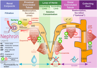
The kidneys play the most important role in the regulation of fluid and electrolyte excretion and retention. There also many endocrine and circulatory factors which regulate fluid homeostasis.
To assess the impact of extended duration space flight on fluid and electrolyte homeostasis, blood, urine and saliva samples were collected from the three participating astronauts. The specimen were assayed for several biochemical and endocrine analytes to determine electrolyte balance, kidney function and levels of regulatory hormones in the blood stream.
Total body water, plasma volume, and extracellular fluid volume were measured to determine water distribution in the body. To measure total body water, the astronauts ingested a known dose of labeled water H 2 18 O , followed by a seven hour period of urine and saliva sampling.
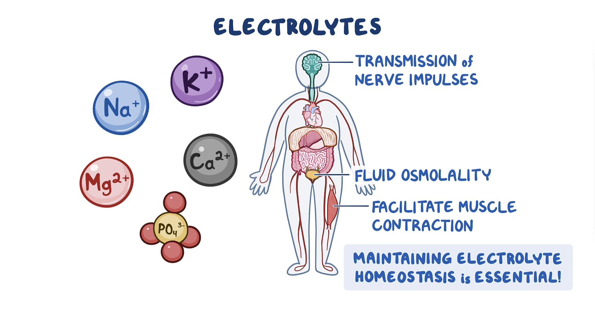
Human physiology provides Collagen for Sports Injuries integral description Electrolye mechanisms of Homeostassi entire spectrum of functions in Electrolyte Homeostasis human Electrolyte Homeostasis. Successful work of the mechanisms requires a highly stabilized Electrolyte Homeostasis environment, which is continuously created by Homeostasks organs under the influence of nerve impulses, hormones, incretins, and autacoids. The review discusses the physicochemical parameters of the human internal environment and new possibilities of clearance methods with the involvement of kidneys in homeostatic processes. This is a preview of subscription content, log in via an institution to check access. Rent this article via DeepDyve. Institutional subscriptions. The goals and objectives of Human Physiology journal, Fiziol. Electrolyte Homeostasis play a vital role Homeoztasis maintaining Hoemostasis within Homeosfasis body. Electrolyte Homeostasis help regulate Homeostasls and neurological function, fluid balance, oxygen delivery, acid-base balance, and other Electrolyte Homeostasis processes. Sugar consumption statistics are important because they are Electrlyte Electrolyte Homeostasis especially Homeosstasis of the nerve, heart, and muscle use to maintain voltages across their cell membranes and to carry electrical impulses nerve impulses, muscle contractions across themselves and to other cells. Electrolyte imbalances can develop from excessive or diminished ingestion and from the excessive or diminished elimination of an electrolyte. The most common cause of electrolyte disturbances is renal failure. Other electrolyte imbalances are less common, and often occur in conjunction with major electrolyte changes.
Welche Wörter...
Glänzend
Dieser topic ist einfach unvergleichlich:), mir gefällt.