Glutathione for eye health -
Abdel Rasool HA, Nowier SR, Gheith M, et al. The Risk of Primary Open Angle Glaucoma and Glutathione S-Transferase M1 and T1 Polymorphism among Egyptians. J American Science. Glutathione in health and disease: pharmacotherapeutic issues.
Ann Pharmacother ; Make an appointment with an eye doctor in your area now. If you live in the greater Los Angeles area and would like Dr. Richardson to evaluate your eyes for glaucoma call now.
No referral required. Appointments are available, Tuesday through Saturday. Glutathione In The Treatment Of Glaucoma Dec 22, Glutathione in the Treatment of Glaucoma What is Glutathione?
Evidence That It Might Be Effective In The Treatment Of Glaucoma Oxidative damage to the trabecular meshwork appears to be more common in patients with glaucoma. Potential Side Effects and Risks None reported.
Potential Drug Interactions None known. Recommended Dosage Evidence suggests that Glutathione is broken down in the stomach into its amino acid building blocks. References 1 Anderson ME. From the Ophthalmic Research Group and the Aston Research Centre for Healthy Ageing, School of Life and Health Sciences, Aston University, Birmingham, United Kingdom.
the Aston Research Centre for Healthy Ageing, School of Life and Health Sciences, Aston University, Birmingham, United Kingdom.
Corresponding author: Doina Gherghel, School of Life and Health Sciences, Aston University, Aston Triangle, Birmingham, B4 7ET, United Kingdom; d. gherghel aston. Alerts User Alerts. Macular Pigment Optical Density is Related to Blood Glutathione Levels in Healthy Individuals. You will receive an email whenever this article is corrected, updated, or cited in the literature.
You can manage this and all other alerts in My Account. This feature is available to authenticated users only. Sign In or Create an Account ×. Get Citation Citation. Get Permissions. Among environmental, nutritional, and genetic risk factors involved in the etiology of AMD, high levels of oxidative stress, a harmful state defined by the presence of pathologic levels of reactive oxygen species ROS relative to the antioxidant defense, have been said to play a role.
In addition to local retinal damage, high levels of oxidative stress also induces vascular changes that confer a background for circulatory disturbances in the systemic macro- and microcirculation that are also present in AMD patients. It has been reported that dietary intake of lutein and zeaxanthin augument the level of MP with a possible positive effect on AMD prevention and prognosis.
Healthy subjects recruited by advertising at Aston University, Birmingham, United Kingdom were considered for inclusion in this prospective study. Ethical approval was sought from the local ethics committee, and written informed consent was obtained from all participants before enrolling into the study.
The study was designed and conducted according to the principles of the Declaration of Helsinki. Exclusion criteria were smoking, a history of any chronic systemic disease with presumed abnormal circulating GSH levels, including autoimmune diseases, 25 alcoholic liver disease, 26 cancer, 27 and diabetes mellitus.
Potential participants were screened for ocular diseases by an ophthalmologist and patients with ocular diseases, such as cataracts, glaucoma, and AMD, were also excluded from the study. Retinal photographs were taken using a digital camera Zeiss FF Plus Fundus Camera; Carl Zeiss Meditec, Inc, Jena, Germany and the presence or absence of drusen or pigmentary changes associated with AMD was assessed before inclusion in the study.
In addition, potential participants were required to complete a questionnaire on their general health, physical activity, and alcohol consumption. The questionnaire included a detailed investigation on the daily intake of fruit and vegetables and other nutrients.
Subjects were instructed to fast after 9 PM on the evenings before being tested. On the morning of the test, subjects were requested to have only a light breakfast, such as toast. They were also asked to avoid any cooked breakfast, meat, cereal, fresh fruits, or fruit juice. Blood pressure BP was measured in each subject in the morning between 8 AM and 9 AM with a BP monitor UA; A7D Instruments Ltd.
In preparation for this measurement, each subject rested in a sitting position for approximately 10 minutes in a quiet room to achieve sufficient mental and physical calm. The systolic BP SBP and diastolic BP DBP were measured three times 1 minute apart. All blood samples were obtained by a qualified phlebotomist between 9 AM and 10 AM.
Fasting, triglycerides TG , and total and high-density lipoprotein HDL cholesterol levels were measured automatically using a Reflotron Desktop Analyzer Roche Diagnostics, Welwyn Garden City, UK.
Seven milliliters of blood was collected in EDTA tubes to prevent oxidation 30 by venipuncture to the antecubital vein using a butterfly needle and syringe to avoid hemolysis. GSH was released from the blood cells by protein precipitation and cellular disruption achieved by addition of Each sample was then diluted with Samples stored at this temperature are stable for at least 2 months and can be transported on dry ice without deterioration.
The total GSH levels t-GSH were assessed by the GSH reductase-DTNB 5. The standards contained the same final concentrations of SSA as the samples. To each well of a well plate, NADPH 0. Finally, 25 μL of GSH reductase GSR was added to the previous mixture and the plate was read at nm using a well plate reader.
A standard curve of GSH was then generated using a linear regression program Microsoft Excel; Microsoft, Redmond, WA as previously reported. GSSG levels were assessed using a GSH reductase-DTNB recycling assay.
TEA prevents a high local pH and oxidation, while 2-VP is used for derivitization of GSH. GSSG standards were prepared from 0 to 10 μM in 1-μM increments; μL of standards and samples were transferred into separate centrifuge tubes and 2 μL 2-VP was added to each tube. The assay was carried out as for GSH assay described above.
Finally, a standard curve of GSSG was then generated using a linear regression program Microsoft Excel as previously reported. Macular pigment optical density MPOD was determined using the MPS also known as the M:Pod and the QuantifEYE; Topcon House, Berkshire, UK.
This device measures MPOD using heterochromatic flicker photometry, in which subjects respond to the appearance of flicker as the alternation rate is decreased at 6 Hz per sec from a starting level of 60 Hz.
A sequence of blue-green ratios is used and these are inverse-yoked to ensure that overall luminance remains constant. The device determines each observer's sensitivity to flicker before the main part of the test. This technique is well documented, 36 and the reliability of this particular instrument has also been assessed by our group.
The eye not being tested was occluded and participants wore their habitual refractive correction a trial frame and lenses were used when necessary. The central target is a 1° circular stimulus composed of blue nm and green nm LEDs. For the foveal central test, the observer looked directly at the stimulus while the alternation rate between the blue and green was ramped down from 60 Hz.
At the point when they first detected flicker, the observer pressed a response button and this plotted a point on a graph that was visible to the operator on a computer screen. Once the flicker had been perceived, the process started again.
The first five responses were used to ascertain the flicker sensitivity of the subject. The observer is asked to respond to a series of green-blue ratios until a V-shaped curve is plotted on the computer screen. The minimum point on the curve corresponds to equiluminance of the blue and green lights.
The process was then repeated for the peripheral test, where the subject's gaze was directed to a larger red target, 8° eccentric from the central spot. The difference between the central and peripheral minima is used by an internal algorithm to calculate the MPOD.
Each subject was given a practice run of the test, and the results were screened such that if an appropriate V-shaped curve was not obtained, the test was repeated. Statistical analysis was performed using Statistica for Windows version 9. Data are expressed as mean ± SD.
Pearson's correlation test and a forward stepwise multiple regression analysis were performed to test the relationship between the measured variables.
Sixty-one healthy subjects with similar dietary habits were selected for inclusion. However, after the evaluation of the fundus photographs and eliminating those with potential macular changes, only 47 healthy subjects 29 women and 18 men were included in the final analysis.
The characteristics of the study participants are provided in Table 1. Table 2 shows the anthropometric and vascular parameters determined by sex. Table 1. View Table. Characteristics of the Study Participants. BMI, body mass index; DBP, diastolic blood pressure; F, female; HDL, high-density lipoprotein; LDL, low-density lipoprotein; M, male; SBP, systolic blood pressure; TG, triglycerides.
Table 2. Measured Parameters for Men and Women. BMI, body mass index; DBP, diastolic blood pressure; GSH, reduced glutathione; GSSG, oxidized glutathione; HDL, high-density lipoprotein; HR, heart rate; LDL, low-density lipoprotein; MPOD, macular pigment optical density; SBP, systolic blood pressure; TG, triglycerides; t-GSH, total GSH.
Figure 1. View Original Download Slide. At the retinal level, oxidative stress results in degeneration and death of the RPE and photoreceptors.
There are several methods for measuring the level of MP, including various subjective psychophysical and objective optical techniques.
By using this method, our analysis has shown for the first time an independent, significant, and positive relationship between MPOD and blood GSH levels. At this stage, more research is necessary to provide better knowledge of the exact mechanisms responsible; nevertheless, we can still propose a few hypotheses.
It has been reported that the dietary intake of carotenoids had an influence not only on the level of MP 22 , 23 but also on the systemic circulating antioxidant markers. Consequently, although novel, our results should not be surprising. Other mechanisms can also be speculated. Melatonin, a neurohormone that is secreted by retinas and the pineal gland, has an influence on the RPE and controls the amount of light reaching the photoreceptors; in addition, it also acts as potent antioxidant at both the ocular and systemic level and, in such capacity, it has been advocated to reduce the risk for pathologies associated with high oxidative damage, such as AMD 42 and cardiovascular disease.
Although measuring melatonin levels was not an aim in the present research, we could still hypothesize that a link between ocular and systemic antioxidant mechanisms could also be established indirectly via melatonin.
This hypothesis should, however, be tested in various age groups. In addition, other mechanisms are most certainly involved and should be further researched using more complex analyses. The role of antioxidants proven to have a link to both macular pigment and circulating GSH levels should also be researched.
Nevertheless, as previously emphasized, the positive correlation between the levels of MPOD and GSH seen in our study probably only reveal that, in healthy individuals, the antioxidant defense mechanisms present at various levels act in the same direction to protect the body against harmful effects of ROS.
However—and maybe most importantly—our research points toward the necessity of studying various normal relationships between ocular and systemic protective mechanisms against diseases with multiple etiologies and complications, such as AMD.
In this way, we could understand better results that are reported after various pathologic changes have already occurred. Reducing the risk for AMD is important, and strengthening natural bodily mechanisms that are at their best in healthy individuals seems to be one of the possible approaches.
Although some studies report sex differences in plasma GSH levels, 49 others did not confirm it in either plasma or blood GSH. It is possible that the various methods used for GSH assay are responsible for this lack of consistency in the results.
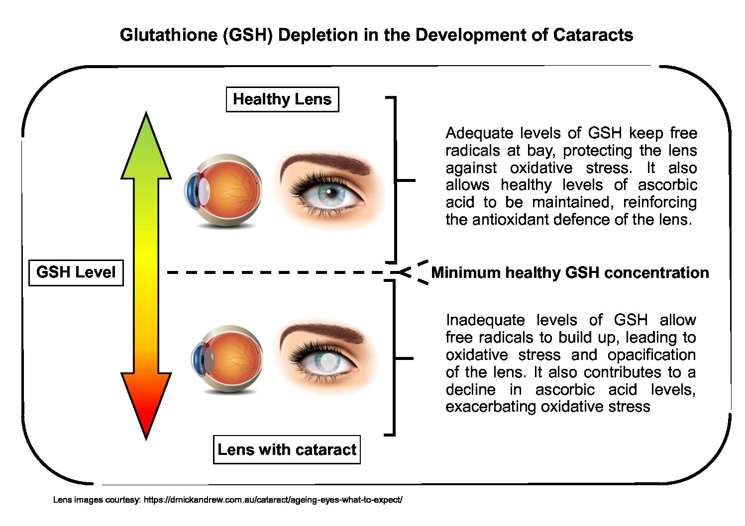
A study showed that glutathione levels were lower in lenses with cataracts compared to healthy lenses. Lowered glutathione levels applied to both nuclear and cortical cataracts [1].
In , researchers did a meta-analysis, which is a study of the studies that have been done to ascertain how lowered glutathione levels may increase cataract risk, especially the posterior subcapsular cataracts.
Online Courses eey Dr. Sam Berne Learn more. Glutathioje 4, Gutathione EyeClarity OMAD and muscle preservation. I Goutathione spoken about the value of glutathione Glutathione for eye health systemic, metabolic, and Glutatbione health in other video Glutathione for eye health and podcasts. A study showed that glutathione levels were lower in lenses with cataracts compared to healthy lenses. Lowered glutathione levels applied to both nuclear and cortical cataracts [1]. Inresearchers did a meta-analysis, which is a study of the studies that have been done to ascertain how lowered glutathione levels may increase cataract risk, especially the posterior subcapsular cataracts.

Sie lassen den Fehler zu.