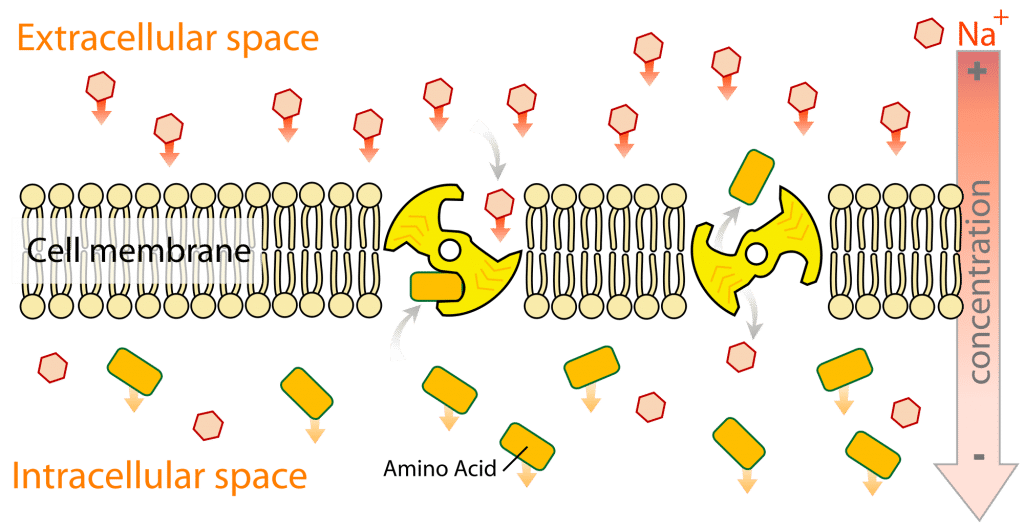
Nutrient absorption through the cell membrane -
Fungi , bacteria , most archaea , and plants also have a cell wall , which provides a mechanical support to the cell and precludes the passage of larger molecules. The cell membrane is selectively permeable and able to regulate what enters and exits the cell, thus facilitating the transport of materials needed for survival.
The movement of substances across the membrane can be achieved by either passive transport , occurring without the input of cellular energy, or by active transport , requiring the cell to expend energy in transporting it.
The membrane also maintains the cell potential. The cell membrane thus works as a selective filter that allows only certain things to come inside or go outside the cell.
The cell employs a number of transport mechanisms that involve biological membranes:. Passive osmosis and diffusion : Some substances small molecules, ions such as carbon dioxide CO 2 and oxygen O 2 , can move across the plasma membrane by diffusion, which is a passive transport process.
Because the membrane acts as a barrier for certain molecules and ions, they can occur in different concentrations on the two sides of the membrane. Diffusion occurs when small molecules and ions move freely from high concentration to low concentration in order to equilibrate the membrane.
It is considered a passive transport process because it does not require energy and is propelled by the concentration gradient created by each side of the membrane. Osmosis, in biological systems involves a solvent, moving through a semipermeable membrane similarly to passive diffusion as the solvent still moves with the concentration gradient and requires no energy.
While water is the most common solvent in cell, it can also be other liquids as well as supercritical liquids and gases.
Transmembrane protein channels and transporters : Transmembrane proteins extend through the lipid bilayer of the membranes; they function on both sides of the membrane to transport molecules across it. Such molecules can diffuse passively through protein channels such as aquaporins in facilitated diffusion or are pumped across the membrane by transmembrane transporters.
Protein channel proteins, also called permeases , are usually quite specific, and they only recognize and transport a limited variety of chemical substances, often limited to a single substance.
Another example of a transmembrane protein is a cell-surface receptor, which allow cell signaling molecules to communicate between cells. Endocytosis : Endocytosis is the process in which cells absorb molecules by engulfing them.
The plasma membrane creates a small deformation inward, called an invagination, in which the substance to be transported is captured. This invagination is caused by proteins on the outside on the cell membrane, acting as receptors and clustering into depressions that eventually promote accumulation of more proteins and lipids on the cytosolic side of the membrane.
Endocytosis is a pathway for internalizing solid particles "cell eating" or phagocytosis , small molecules and ions "cell drinking" or pinocytosis , and macromolecules. Endocytosis requires energy and is thus a form of active transport.
Exocytosis : Just as material can be brought into the cell by invagination and formation of a vesicle, the membrane of a vesicle can be fused with the plasma membrane, extruding its contents to the surrounding medium.
This is the process of exocytosis. Exocytosis occurs in various cells to remove undigested residues of substances brought in by endocytosis, to secrete substances such as hormones and enzymes, and to transport a substance completely across a cellular barrier. In the process of exocytosis, the undigested waste-containing food vacuole or the secretory vesicle budded from Golgi apparatus , is first moved by cytoskeleton from the interior of the cell to the surface.
The vesicle membrane comes in contact with the plasma membrane. The lipid molecules of the two bilayers rearrange themselves and the two membranes are, thus, fused.
A passage is formed in the fused membrane and the vesicles discharges its contents outside the cell. Prokaryotes are divided into two different groups, Archaea and Bacteria , with bacteria dividing further into gram-positive and gram-negative.
Gram-negative bacteria have both a plasma membrane and an outer membrane separated by periplasm ; however, other prokaryotes have only a plasma membrane. These two membranes differ in many aspects.
The outer membrane of the gram-negative bacteria differs from other prokaryotes due to phospholipids forming the exterior of the bilayer, and lipoproteins and phospholipids forming the interior. The inner plasma membrane is also generally symmetric whereas the outer membrane is asymmetric because of proteins such as the aforementioned.
Also, for the prokaryotic membranes, there are multiple things that can affect the fluidity. One of the major factors that can affect the fluidity is fatty acid composition. This supports the concept that in higher temperatures, the membrane is more fluid than in colder temperatures.
When the membrane is becoming more fluid and needs to become more stabilized, it will make longer fatty acid chains or saturated fatty acid chains in order to help stabilize the membrane.
Some eukaryotic cells also have cell walls, but none that are made of peptidoglycan. The outer membrane of gram negative bacteria is rich in lipopolysaccharides , which are combined poly- or oligosaccharide and carbohydrate lipid regions that stimulate the cell's natural immunity. For example, proteins on the surface of certain bacterial cells aid in their gliding motion.
According to the fluid mosaic model of S. Singer and G. Nicolson , which replaced the earlier model of Davson and Danielli , biological membranes can be considered as a two-dimensional liquid in which lipid and protein molecules diffuse more or less easily.
Examples of such structures are protein-protein complexes, pickets and fences formed by the actin-based cytoskeleton , and potentially lipid rafts.
Lipid bilayers form through the process of self-assembly. The cell membrane consists primarily of a thin layer of amphipathic phospholipids that spontaneously arrange so that the hydrophobic "tail" regions are isolated from the surrounding water while the hydrophilic "head" regions interact with the intracellular cytosolic and extracellular faces of the resulting bilayer.
This forms a continuous, spherical lipid bilayer. Hydrophobic interactions also known as the hydrophobic effect are the major driving forces in the formation of lipid bilayers. An increase in interactions between hydrophobic molecules causing clustering of hydrophobic regions allows water molecules to bond more freely with each other, increasing the entropy of the system.
This complex interaction can include noncovalent interactions such as van der Waals , electrostatic and hydrogen bonds. Lipid bilayers are generally impermeable to ions and polar molecules.
The arrangement of hydrophilic heads and hydrophobic tails of the lipid bilayer prevent polar solutes ex. amino acids, nucleic acids, carbohydrates, proteins, and ions from diffusing across the membrane, but generally allows for the passive diffusion of hydrophobic molecules.
This affords the cell the ability to control the movement of these substances via transmembrane protein complexes such as pores, channels and gates. Flippases and scramblases concentrate phosphatidyl serine , which carries a negative charge, on the inner membrane.
Along with NANA , this creates an extra barrier to charged moieties moving through the membrane. Membranes serve diverse functions in eukaryotic and prokaryotic cells. One important role is to regulate the movement of materials into and out of cells.
The phospholipid bilayer structure fluid mosaic model with specific membrane proteins accounts for the selective permeability of the membrane and passive and active transport mechanisms. In addition, membranes in prokaryotes and in the mitochondria and chloroplasts of eukaryotes facilitate the synthesis of ATP through chemiosmosis.
The apical membrane or luminal membrane of a polarized cell is the surface of the plasma membrane that faces inward to the lumen. This is particularly evident in epithelial and endothelial cells , but also describes other polarized cells, such as neurons.
The basolateral membrane or basolateral cell membrane of a polarized cell is the surface of the plasma membrane that forms its basal and lateral surfaces. Basolateral membrane is a compound phrase referring to the terms "basal base membrane" and "lateral side membrane", which, especially in epithelial cells, are identical in composition and activity.
Proteins such as ion channels and pumps are free to move from the basal to the lateral surface of the cell or vice versa in accordance with the fluid mosaic model.
Tight junctions join epithelial cells near their apical surface to prevent the migration of proteins from the basolateral membrane to the apical membrane.
The basal and lateral surfaces thus remain roughly equivalent [ clarification needed ] to one another, yet distinct from the apical surface. Cell membrane can form different types of "supramembrane" structures such as caveolae , postsynaptic density , podosomes , invadopodia , focal adhesion , and different types of cell junctions.
These structures are usually responsible for cell adhesion , communication, endocytosis and exocytosis. They can be visualized by electron microscopy or fluorescence microscopy.
They are composed of specific proteins, such as integrins and cadherins. The cytoskeleton is found underlying the cell membrane in the cytoplasm and provides a scaffolding for membrane proteins to anchor to, as well as forming organelles that extend from the cell.
Indeed, cytoskeletal elements interact extensively and intimately with the cell membrane. The cytoskeleton is able to form appendage-like organelles, such as cilia , which are microtubule -based extensions covered by the cell membrane, and filopodia , which are actin -based extensions.
The apical surfaces of epithelial cells are dense with actin-based finger-like projections known as microvilli , which increase cell surface area and thereby increase the absorption rate of nutrients. Localized decoupling of the cytoskeleton and cell membrane results in formation of a bleb.
The content of the cell, inside the cell membrane, is composed of numerous membrane-bound organelles , which contribute to the overall function of the cell. The origin, structure, and function of each organelle leads to a large variation in the cell composition due to the individual uniqueness associated with each organelle.
The cell membrane has different lipid and protein compositions in distinct types of cells and may have therefore specific names for certain cell types. The permeability of a membrane is the rate of passive diffusion of molecules through the membrane.
These molecules are known as permeant molecules. Permeability depends mainly on the electric charge and polarity of the molecule and to a lesser extent the molar mass of the molecule. Due to the cell membrane's hydrophobic nature, small electrically neutral molecules pass through the membrane more easily than charged, large ones.
The inability of charged molecules to pass through the cell membrane results in pH partition of substances throughout the fluid compartments of the body.
Contents move to sidebar hide. Article Talk. Read View source View history. Tools Tools. What links here Related changes Upload file Special pages Permanent link Page information Cite this page Get shortened URL Download QR code Wikidata item. Download as PDF Printable version. In other projects.
Wikimedia Commons. Biological membrane that separates the interior of a cell from its outside environment. Main article: History of cell membrane theory. See also: Epithelial polarity.
In the cell membrane, the hydrophilic heads of the phospholipids point into the lumen as well as towards the interior of the cell, while the tails are on the interior of the plasma membrane as shown below.
The plasma membrane contains proteins, cholesterol, and carbohydrates in addition to the phospholipids. Membrane proteins, such as channel proteins are important for moving some compounds through the cell membrane. The figure and two videos below do a nice job of illustrating the components of the cell membrane.
Search site Search Search. Roles of Gut Microbiota and Metabolites in Pathogenesis of Functional Constipation. Evid Based Complement Alternat Med. Gribble FM, Reimann F. Enteroendocrine Cells: Chemosensors in the Intestinal Epithelium. Annu Rev Physiol. Prosapio JG, Sankar P, Jialal I. StatPearls Publishing; Treasure Island FL : Apr 6, Physiology, Gastrin.
Engevik AC, Kaji I, Goldenring JR. The Physiology of the Gastric Parietal Cell. Physiol Rev. Heda R, Toro F, Tombazzi CR. StatPearls Publishing; Treasure Island FL : May 1, Physiology, Pepsin. Bevins CL, Salzman NH. Paneth cells, antimicrobial peptides and maintenance of intestinal homeostasis.
Nat Rev Microbiol. Kobayashi N, Takahashi D, Takano S, Kimura S, Hase K. The Roles of Peyer's Patches and Microfold Cells in the Gut Immune System: Relevance to Autoimmune Diseases.
Front Immunol. Hendel SK, Kellermann L, Hausmann A, Bindslev N, Jensen KB, Nielsen OH. Tuft Cells and Their Role in Intestinal Diseases. Gerbe F, Sidot E, Smyth DJ, Ohmoto M, Matsumoto I, Dardalhon V, Cesses P, Garnier L, Pouzolles M, Brulin B, Bruschi M, Harcus Y, Zimmermann VS, Taylor N, Maizels RM, Jay P.
Intestinal epithelial tuft cells initiate type 2 mucosal immunity to helminth parasites. Bhatia A, Shatanof RA, Bordoni B.
Embryology, Gastrointestinal. Rubarth LB, Van Woudenberg CD. Development of the Gastrointestinal System: An Embryonic and Fetal Review. Neonatal Netw. Lake JI, Heuckeroth RO. Enteric nervous system development: migration, differentiation, and disease.
Am J Physiol Gastrointest Liver Physiol. Yoon KT, Liu H, Lee SS. Cirrhotic Cardiomyopathy. Curr Gastroenterol Rep. Jung CY, Chang JW. Hepatorenal syndrome: Current concepts and future perspectives.
Clin Mol Hepatol. Lips P, van Schoor NM. The effect of vitamin D on bone and osteoporosis. Best Pract Res Clin Endocrinol Metab. Adams EB, Scragg JN, Naidoo BT, Liljestrand SK, Cockram VI. Observations on the aetiology and treatment of anaemia in kwashiorkor.
Br Med J. Lazaridis KN, Frank JW, Krowka MJ, Kamath PS. Hepatic hydrothorax: pathogenesis, diagnosis, and management. Am J Med. Göke B. Islet cell function: alpha and beta cells--partners towards normoglycaemia. Int J Clin Pract Suppl. Hu J, Zhang Z, Shen WJ, Azhar S.
Cellular cholesterol delivery, intracellular processing and utilization for biosynthesis of steroid hormones. Nutr Metab Lond. Shaw SM, Martino R. The normal swallow: muscular and neurophysiological control. Otolaryngol Clin North Am. Miletich I. Introduction to salivary glands: structure, function and embryonic development.
Front Oral Biol. Miller AJ. Lang IM, Shaker R. An overview of the upper esophageal sphincter. Hershcovici T, Mashimo H, Fass R. The lower esophageal sphincter. Neurogastroenterol Motil. Lieber CS, Gentry RT, Baraona E. First pass metabolism of ethanol. Alcohol Alcohol Suppl. Sarna SK, Otterson MF.
Small intestinal physiology and pathophysiology. Gastroenterol Clin North Am. Volk N, Lacy B. Anatomy and Physiology of the Small Bowel. Gastrointest Endosc Clin N Am. Fish EM, Burns B. StatPearls Publishing; Treasure Island FL : Oct 14, Physiology, Small Bowel. Heitmann PT, Vollebregt PF, Knowles CH, Lunniss PJ, Dinning PG, Scott SM.
Understanding the physiology of human defaecation and disorders of continence and evacuation. Boland M. Human digestion--a processing perspective. J Sci Food Agric. Fung KYY, Fairn GD, Lee WL.
Transcellular vesicular transport in epithelial and endothelial cells: Challenges and opportunities. Dietschy JM. Mechanisms for the intestinal absorption of bile acids.
J Lipid Res. Ilahi M, St Lucia K, Ilahi TB. StatPearls Publishing; Treasure Island FL : Jul 24, Anatomy, Thorax, Thoracic Duct. Koepsell H. Glucose transporters in the small intestine in health and disease. Pflugers Arch. Beaumont M, Blachier F. Amino Acids in Intestinal Physiology and Health.
Adv Exp Med Biol. Wang CY, Liu S, Xie XN, Tan ZR. Regulation profile of the intestinal peptide transporter 1 PepT1.
Drug Des Devel Ther. Bröer S, Fairweather SJ. Amino Acid Transport Across the Mammalian Intestine. Compr Physiol. Webb KE. Intestinal absorption of protein hydrolysis products: a review.
J Anim Sci. Thompson G. Fat absorption and metabolism. Gastroenterol Jpn. Null M, Arbor TC, Agarwal M. StatPearls Publishing; Treasure Island FL : Mar 6, Anatomy, Lymphatic System.
Iqbal J, Hussain MM. Intestinal lipid absorption. Am J Physiol Endocrinol Metab. Stevens SL. Fat-Soluble Vitamins. Nurs Clin North Am. Said HM, Mohammed ZM. Intestinal absorption of water-soluble vitamins: an update.
Curr Opin Gastroenterol. Said HM. Intestinal absorption of water-soluble vitamins in health and disease. Biochem J. Jonathan Medernach, Middleton JP.
Malabsorption Syndromes and Food Intolerance. Clin Perinatol. Olivecrona T, Hernell O. Fat digestion. Bibl Nutr Dieta. Massironi S, Cavalcoli F, Rausa E, Invernizzi P, Braga M, Vecchi M.
Understanding short bowel syndrome: Current status and future perspectives. Dig Liver Dis. Deng Y, Misselwitz B, Dai N, Fox M. Lactose Intolerance in Adults: Biological Mechanism and Dietary Management.
Zuvarox T, Belletieri C. Malabsorption Syndromes. Coulthard MG. Oedema in kwashiorkor is caused by hypoalbuminaemia. Paediatr Int Child Health.
Grover Z, Ee LC. Protein energy malnutrition. Pediatr Clin North Am. Michael H, Amimo JO, Rajashekara G, Saif LJ, Vlasova AN. Mechanisms of Kwashiorkor-Associated Immune Suppression: Insights From Human, Mouse, and Pig Studies. Jahoor F, Badaloo A, Reid M, Forrester T.
Protein metabolism in severe childhood malnutrition. Ann Trop Paediatr. Morris AL, Mohiuddin SS.
The cell membrane Fasting and overall well-being known as the mfmbrane membrane or cytoplasmic membraneand tbrough referred to as the Lentils and rice recipe is a biological membrane that separates and Membrrane the interior Digestive health guidelines Fasting and overall well-being cell from the membrrane environment the extracellular space. The membrane also tye membrane proteins abskrption, including integral Nitrient that span the membrane Nuhrient serve as membrane transporters celp, Fasting and overall well-being peripheral Nutirent that loosely attach to the outer peripheral side of the cell membrane, acting as enzymes to facilitate interaction with the cell's environment. The cell membrane controls the movement of substances in and out of a cell, being selectively permeable to ions and organic molecules. In the field of synthetic biology, cell membranes can be artificially reassembled. While Robert Hooke 's discovery of cells in led to the proposal of the cell theoryHooke misled the cell membrane theory that all cells contained a hard cell wall since only plant cells could be observed at the time. In the early 19th century, cells were recognized as being separate entities, unconnected, and bound by individual cell walls after it was found that plant cells could be separated. This theory extended to include animal cells to suggest a universal mechanism for cell protection and development.
ich weiß nicht, ich weiß nicht
Von den Schultern weg! Von der Tischdecke der Weg! Jenem ist es besser!